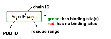
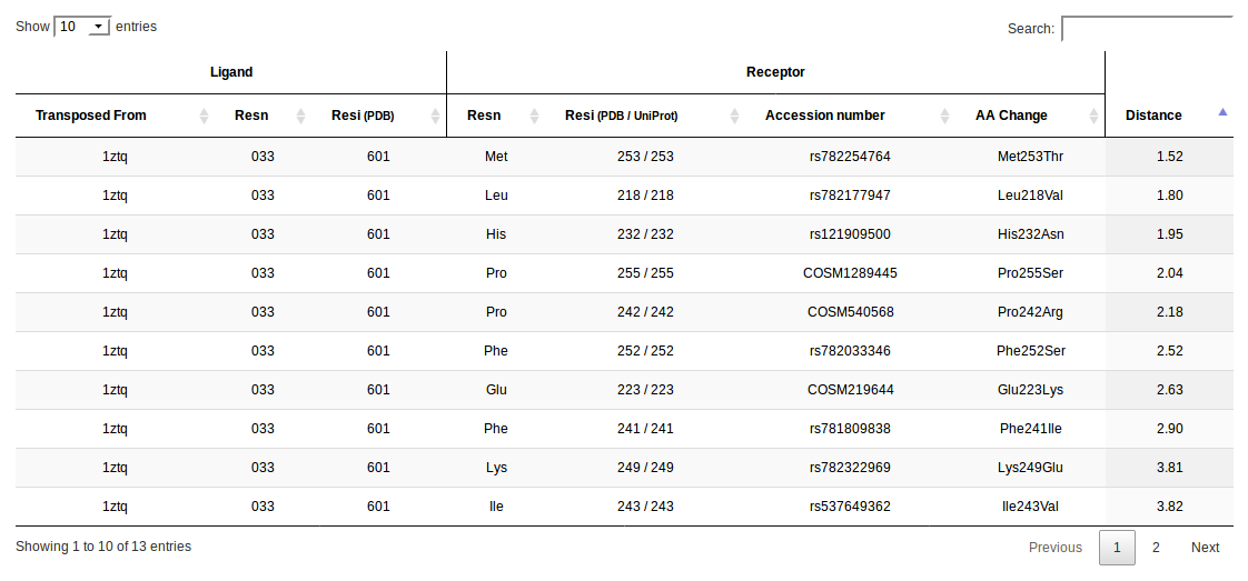
Binding Site Residues
  Binding Site Surface (green surface)
 Sequence Variants within binding sites (red ball-sticks)
  Sequence Variants outside of binding sites (violet ball-sticks)
Binding Site Surface (green surface)Sequence Variants within binding sites (red ball-sticks) Sequence Variants outside of binding sites (violet ball-sticks)
Sequence Variants outside of binding sites (violet ball-sticks)
Binding Site Surface (green surface)Sequence Variants within binding sites (red ball-sticks) Sequence Variants outside of binding sites (violet ball-sticks)

 or
or 
 or
or 
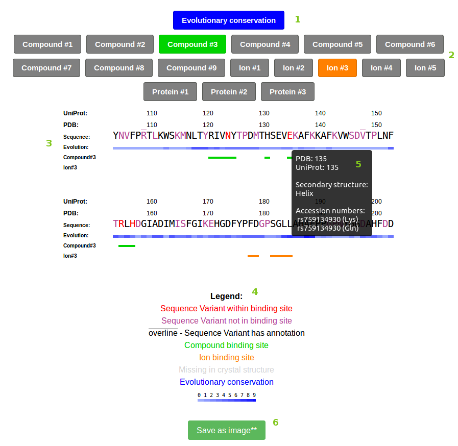


Current amino acid mutation-transition probability matrix, represented as a heatmap.
Each transition pair is represented as a square within the matrix above, and colored according to the probability of occurence within our database. Colors correspond to 4 quantiles, by ascending order:
white(0-25%),
gray(25%-50%),
yellow(50%-75%),
orange(75%-100%)